
Synapses Anatomy and Physiology I
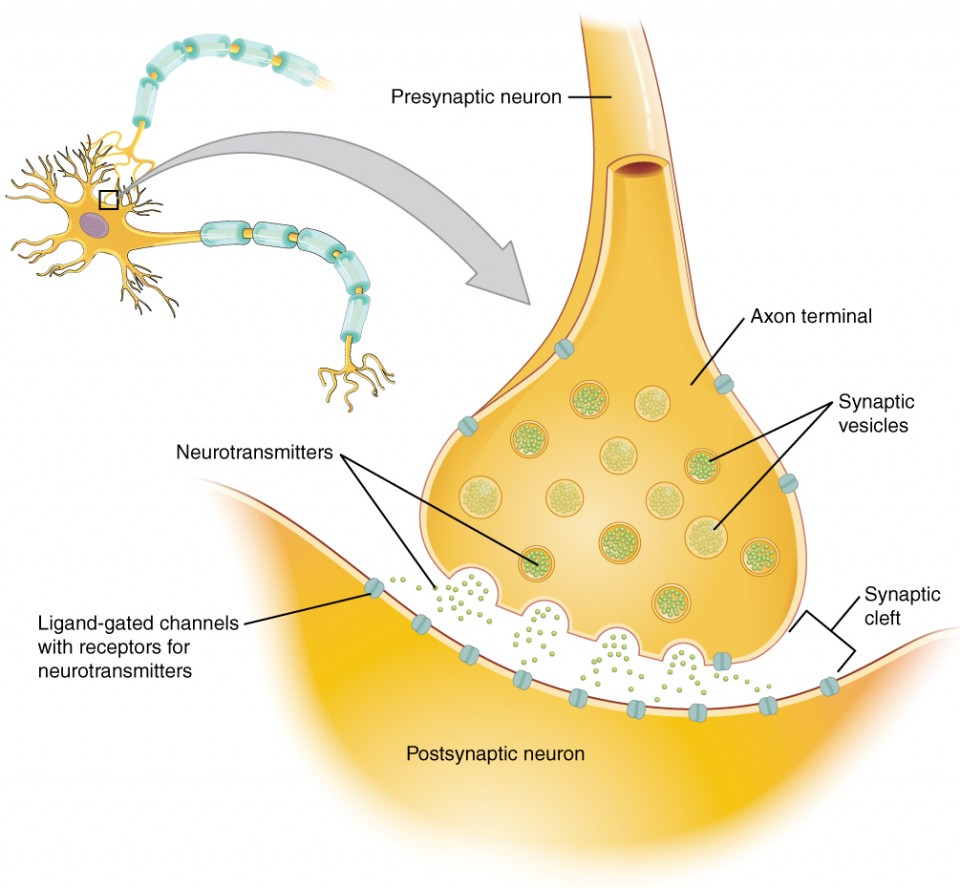
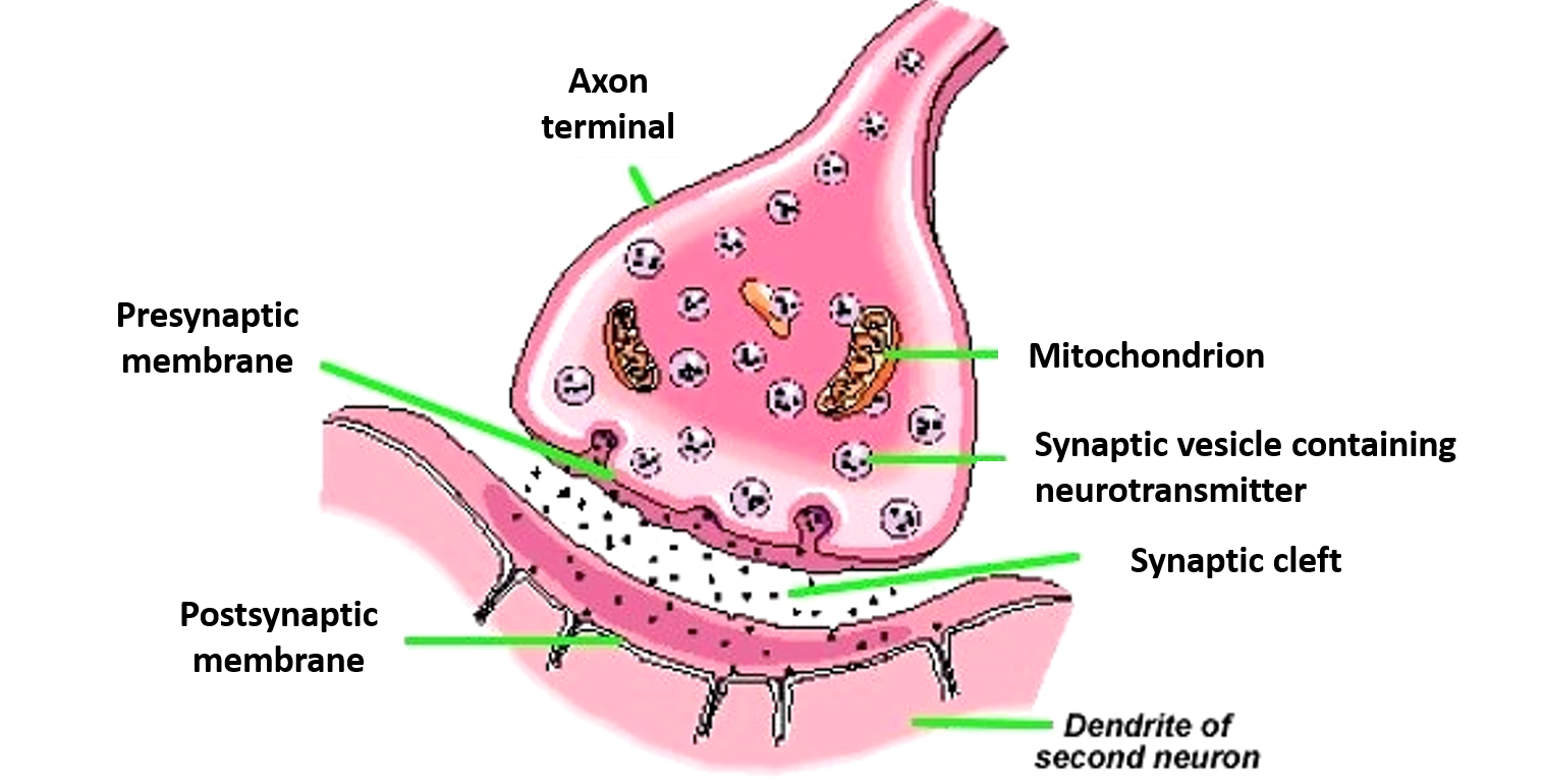
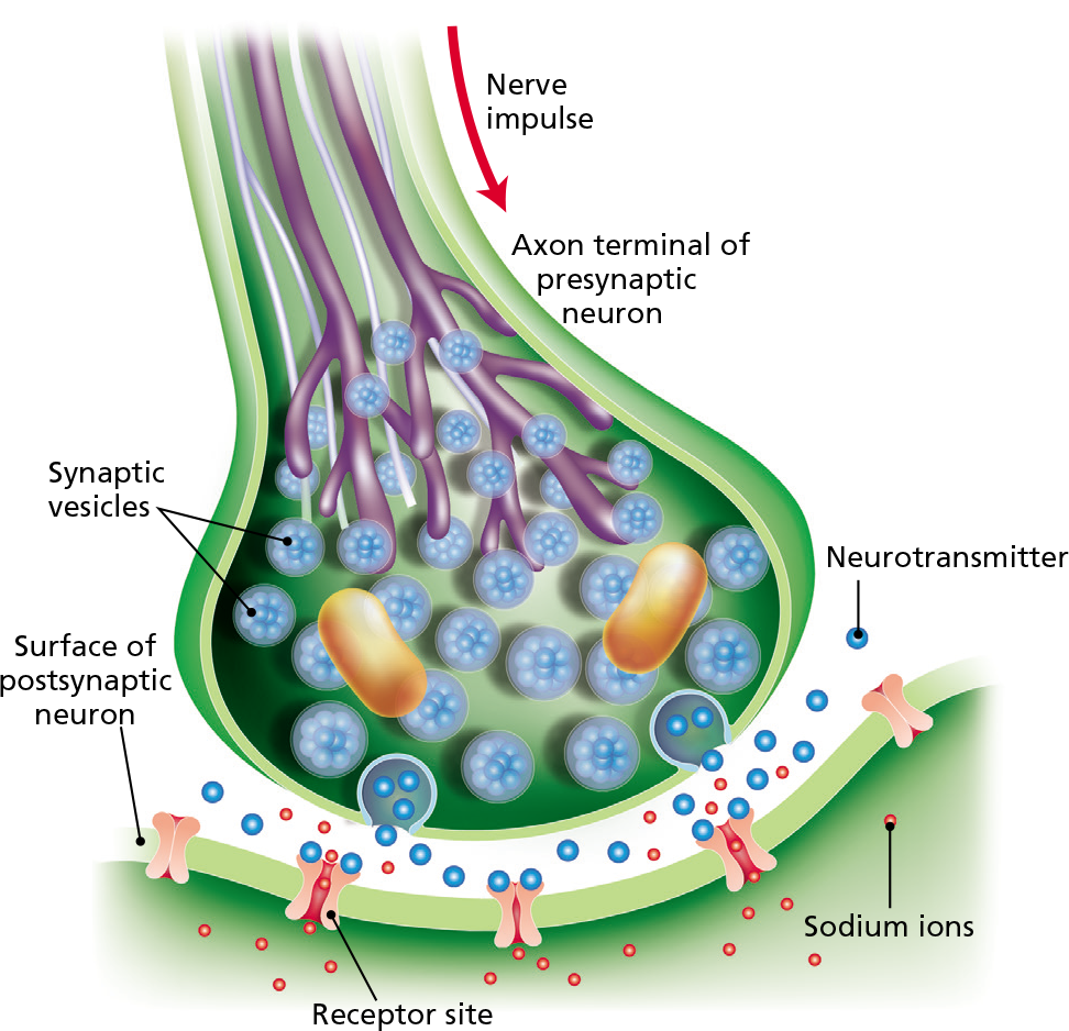
Diagram of a synapse, showing neurotransmitters stored in synaptic vesicles inside the axon terminal. In response to an action potential, the vesicles fuse with the presynaptic membrane and release neurotransmitter into the synaptic cleft. Image modified from " The synapse ," by OpenStax College, Anatomy & Physiology ( CC BY 3.0 ).
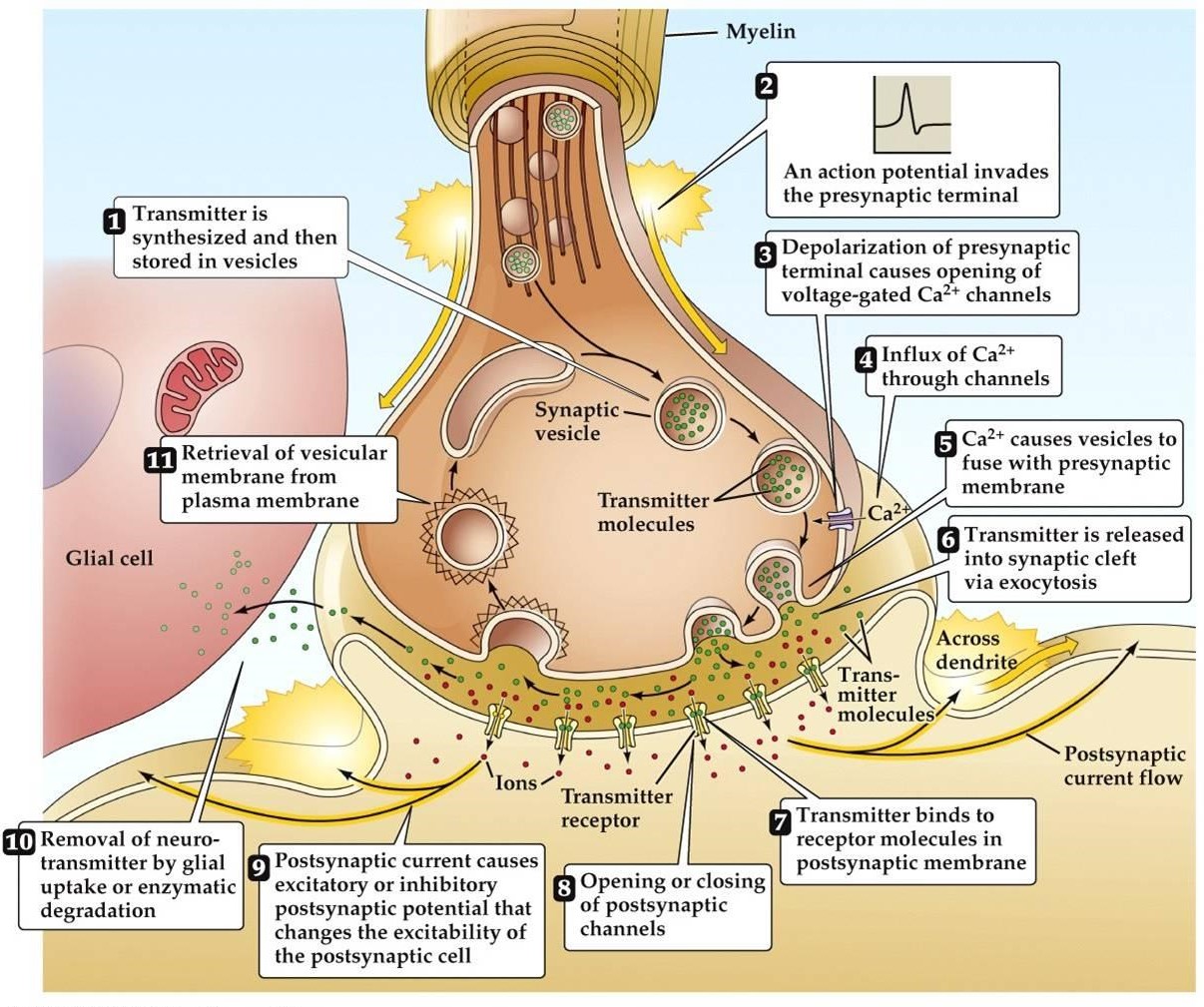
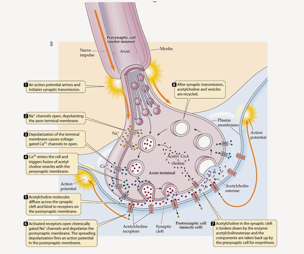
Synaptic transmission steps, Synapses types and Nature of the postsynaptic change Science online
Resources. For the nervous system to function, neurons must be able to communicate with each other, and they do this through structures called synapses. At the synapse, the terminal of a presynaptic cell comes into close contact with the cell membrane of a postsynaptic neuron. Figure 8.1. The terminal of a presynaptic neuron comes into close.

Chemical and Electrical Synapses Biology for Majors II
At its simplest, the neuromuscular junction is a type of synapse where neuronal signals from the brain or spinal cord interact with skeletal muscle fibers, causing them to contract. The activation of many muscle fibers together causes muscles to contract, which in turn can produce movement.
Introduction to Neurophysiology Physiopedia
Where two neurons meet there is a small gap called a synapse close synapse A tiny gap at the junction between two nerve cells, which nerve signals must cross..The plasma membranes of each neuron.
Microglia Maintenance of Neuron Synapses
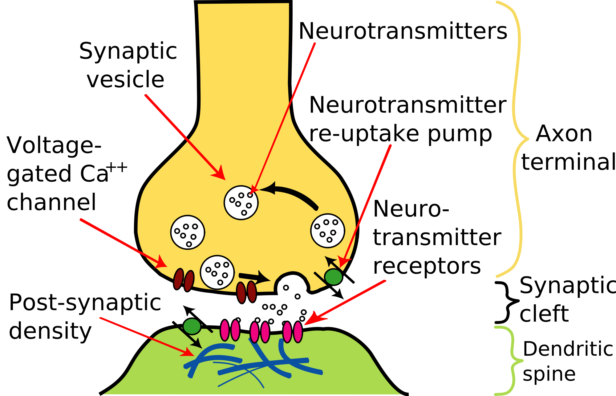
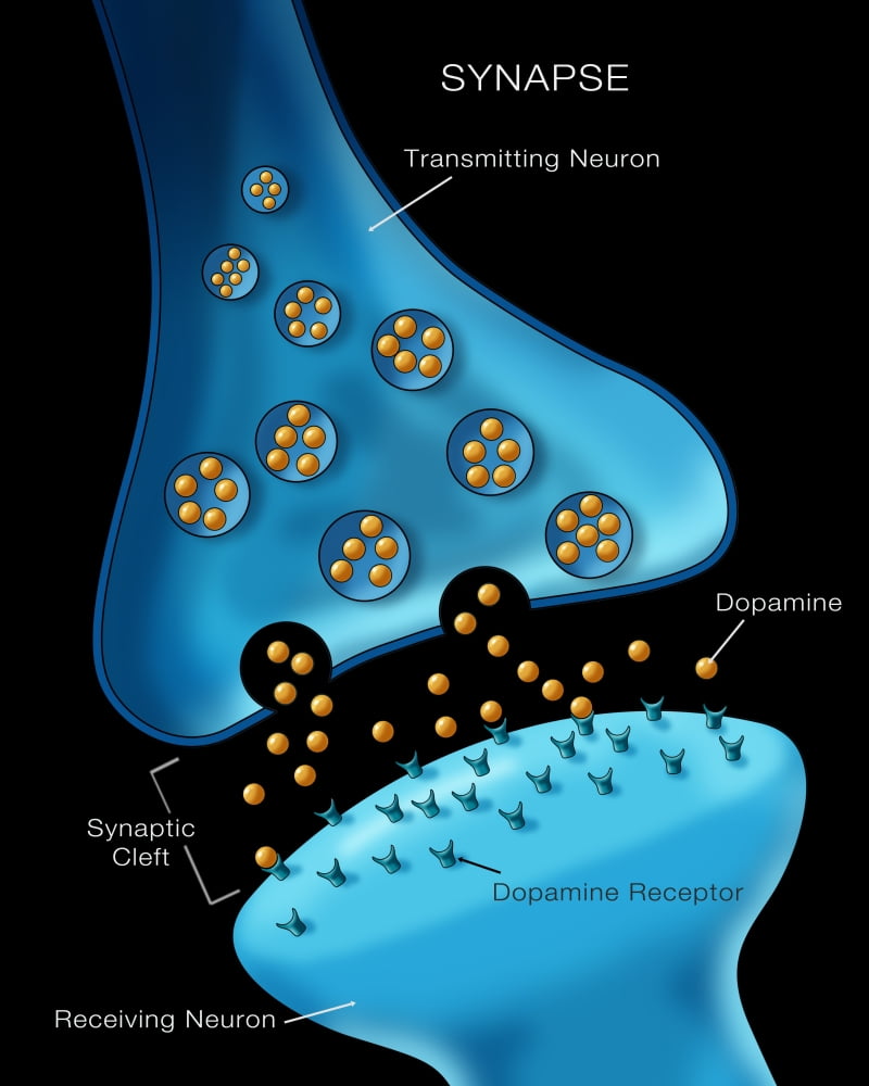
Types Synapses are fundamentally classified into two different types based on how the neurons function to communicate: 1) chemical synapse and 2) electrical synapse. 1) Chemical Synapse Found in vertebrates, it works using neurotransmitters that establish the virtual connection between the presynaptic and postsynaptic neurons.
Communication Between Neurons Anatomy and Physiology I
¡Precios increíbles y alta calidad aquí en Temu. Envío gratuito en todos los pedidos. ¡Solo hoy, disfruta de todas las categorías hasta un 90% de descuento en tu compra.
Anatomy synapse cells transmission signal Vector Image
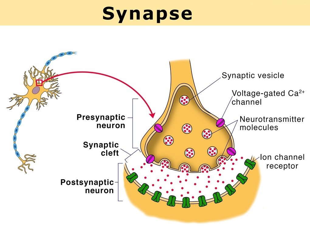
The general structure of a chemical synapse is shown schematically in Figure 5.1B. The space between the pre- and postsynaptic neurons is substantially greater at chemical synapses than at electrical synapses and is called the synaptic cleft. However, the key feature of all chemical synapses is the presence of small, membrane-bounded organelles called synaptic vesicles within the presynaptic.

2 A schematic representation of synapse. Download Scientific Diagram
The peripheral nervous system ( PNS ), which consists of the neurons and parts of neurons found outside of the CNS, includes sensory neurons and motor neurons. Sensory neurons bring signals into the CNS, and motor neurons carry signals out of the CNS. _Image modified from " Nervous system diagram ," by Medium69 ( CC BY-SA 4.0 )._
Synapses Definition, Types and Structure , Anatomy QA
synapse, the site of transmission of electric nerve impulses between two nerve cells (neurons) or between a neuron and a gland or muscle cell (effector). A synaptic connection between a neuron and a muscle cell is called a neuromuscular junction.
Biology learnspot The Synapse and mechanism of synaptic transmission.
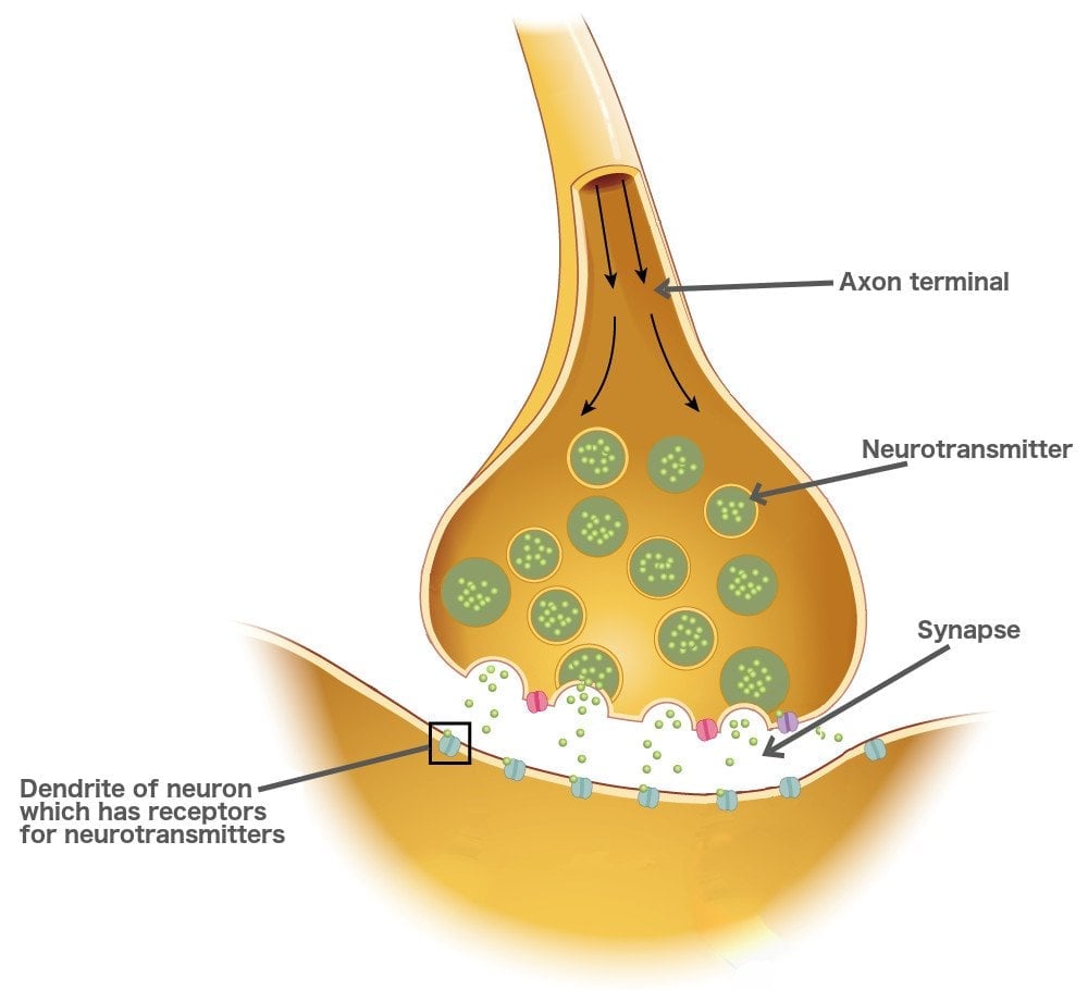
Diagram of a chemical synaptic connection. In the nervous system, a synapse [1] is a structure that permits a neuron (or nerve cell) to pass an electrical or chemical signal to another neuron or to the target effector cell.

What Happens at The Synapse?
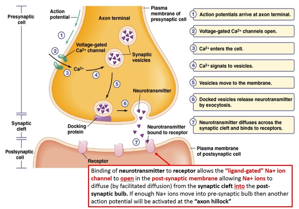
At the synapse, the firing of an action potential in one neuron—the presynaptic, or sending, neuron—causes the transmission of a signal to another neuron—the postsynaptic, or receiving, neuron—making the postsynaptic neuron either more or less likely to fire its own action potential.
Synapses
Mechanism of Synaptic Transmission 3. Properties. Definition of Synapse: Synapse can be defined as functional junction between parts of two different neurons. There is no anatomical continuity between two neurons involved in the formation of synapse.
Neuron Synapse Anatomy Poster Print by Monica Schroeder/Science Source (24 x 36)
Synapse diagram Each neuron forms about 2,000 synapses. There are about 10 11 neurons in the CNS. The synapses are of different types and can be classified on the following bases. Parts of neurons involved in the synapse Axodendritic synapse- The axon of the presynaptic neuron connects to the dendrite of the postsynaptic neuron.

representative synapse Pearson education, Nerve cell, Cell diagram
A synapse is a small gap at the end of a neuron that allows a signal to pass from one neuron to the next. Neurons are cells that transmit information between your brain and other parts of the central nervous system. Synapses are found where neurons connect with other neurons.
Neurotransmission
In psychology, a synapse is the junction between two neurons where information is transmitted from one neuron to another. It consists of the axon terminal of the sending neuron, the synaptic cleft (a small gap), and the dendrite or cell body of the receiving neuron.
What Is A Synapse? What Is A Neurotransmitter? How Do They Work?
(A) Electron micrograph of a human synapse with two synaptic junctions to illustrate the canonical features of all synapses: An intercellular junction in which a presynaptic varicosity that is filled with synaptic vesicles contacts a postsynaptic dendrite that contains multiple trafficking organelles as well as ribosomes (image courtesy of Dr. C.
