
Interior View of the Inferior Skull
A short lecture by Dr. Kathleen Alsup introducing students to the anatomy of the skull from an inferior view.Check out our website (LINK BELOW) for additiona.
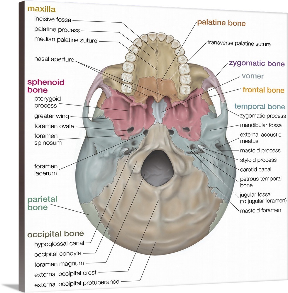
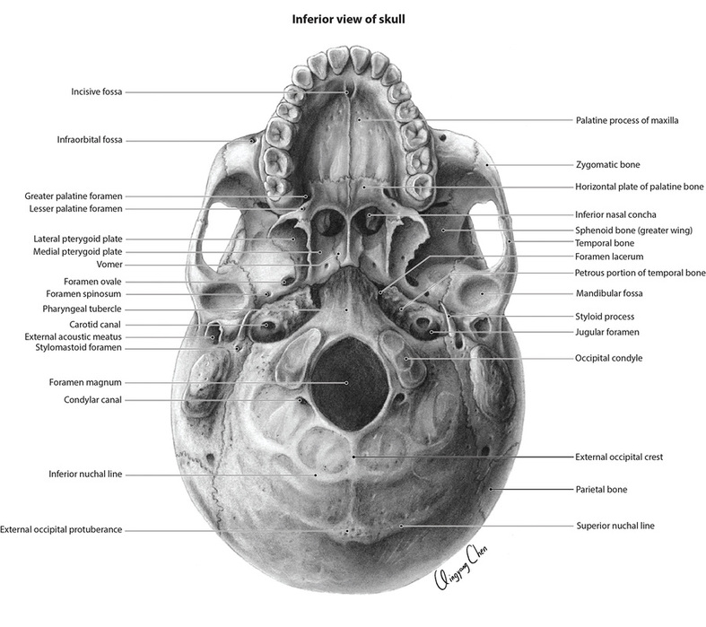
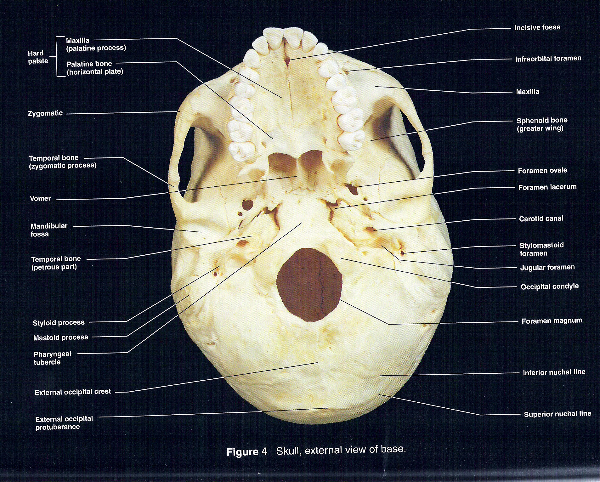
Skull inferior view. skeletal system Wall Art, Canvas Prints, Framed
1/2 Synonyms: none The human skull consists of 22 bones (or 29, including the inner ear bones and hyoid bone) which are mostly connected together by ossified joints, so called sutures. The skull is divided into the braincase ( neurocr anium) and the facial skeleton ( viscerocranium ).
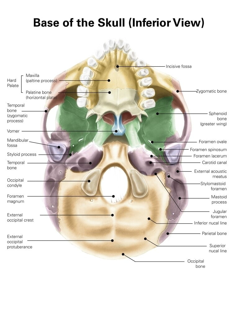
Colored base of human skull, inferior view, with labels. Poster Print
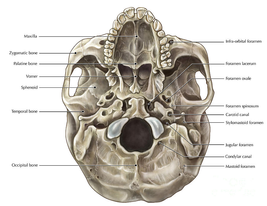
A better view of the vomer bone is seen when looking into the posterior nasal cavity with an inferior view of the skull, where the vomer forms the full height of the nasal septum. The anterior nasal septum is formed by the septal cartilage, a flexible plate that fills in the gap between the perpendicular plate of the ethmoid and vomer bones.

Inferior view of the skull Body bones, Free education, Occipital
These processes comprise lateral and medial plates; the most inferior "hook" Mandible, Top anterior view, middle posterior view, bottom lateral view. of the medial plate is called the hamulus of the pterygoid. Additionally, the medial plate comprises part of the nasal walls. Perforating the root of the large wing inferiorly are three foramina.
QCVisual Portfolio Inferior View of the Skull QCVisual
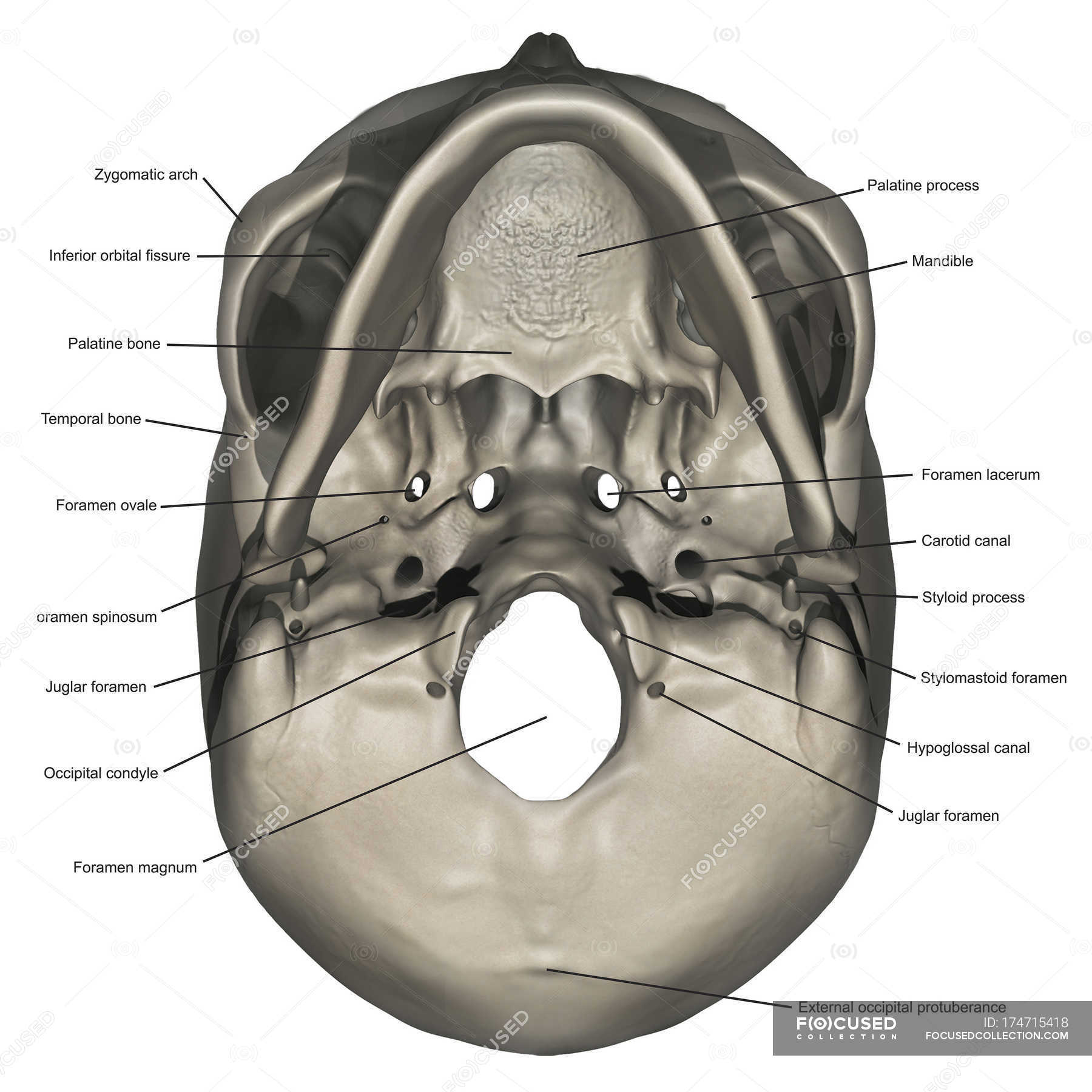
Structures seen on the inferior view of the base of the skull. This article will describe the anatomy from the inferior view of the skull base. We will explore the many foramina and projections that enable arteries and nerves to both enter and leave the skull.
Inferior View of Skull
A better view of the vomer bone is seen when looking into the posterior nasal cavity with an inferior view of the skull, where the vomer forms the full height of the nasal septum. The anterior nasal septum is formed by the septal cartilage, a flexible plate that fills in the gap between the perpendicular plate of the ethmoid and vomer bones.

The inferior view of the adult skull. (With images) Anatomy bones
This is the last view to be discussed in the anatomical views of skull. This inferior view of skull includes: - Bones- Foramina & canals- Fissures, lines & g.
Inferior View Of Skull Skull Anatomy Anatomy Bones Anatomy My XXX Hot
Inferior View of the Base of the Skull (preview) - Human Anatomy | Kenhub Kenhub - Learn Human Anatomy 1.17M subscribers Subscribe 322 From a channel with a licensed health professional in.

The Skull Anatomy and Physiology I
inferior view of the human skull In humans the base of the cranium is the occipital bone, which has a central opening ( foramen magnum) to admit the spinal cord.
Skull Inferior View Photograph by Evan Oto Fine Art America
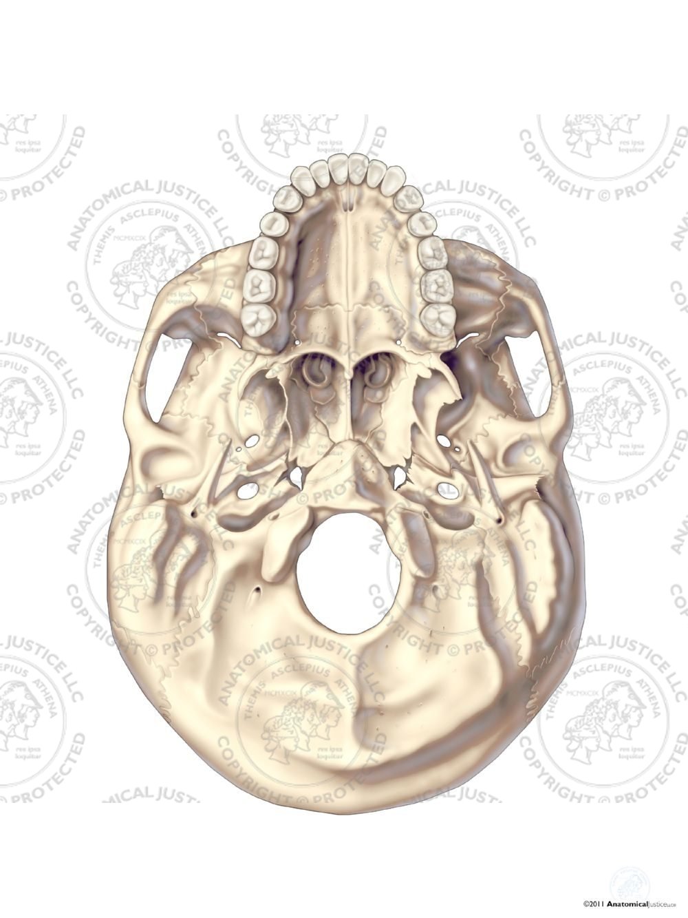
Vomer. Inferior view of the base of the skull. The foramen magnum is the largest foramen on the skull base, through which the spinal cord enters the cranium. The occipital condyles occupy the anterolateral aspects of the foramen magnum and are the site of articulation with the cervical atlas. Prominent foramina visible here for intracranial.

The Skull Anatomy and Physiology
Watch this video to view a rotating and exploded skull, with color-coded bones. Which bone (yellow) is centrally located and joins with most of the other bones of the skull? Anterior View of Skull
Principles of Human Anatomy and Physiology CHAPTER 7 Anatomy of Bones
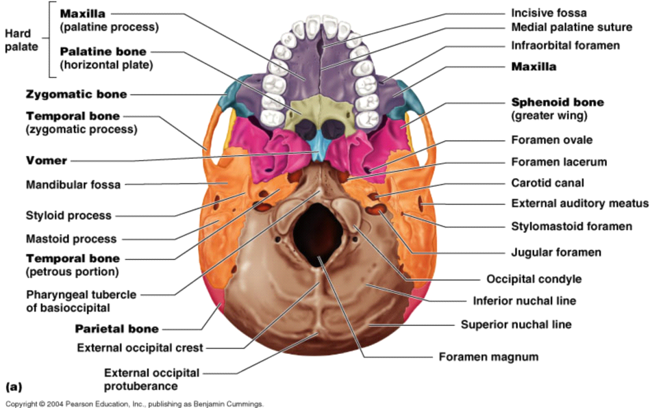
Cranial bones of the skull - inferior view 1 2 3 4 5 Maxilla Bone: Palatine process of maxilla ( processus palatinus maxillae ). Incisive foramen ( foramen incisivum maxillae ). Markings of the maxilla bone - inferior view 1
Skull cranial base, inferior view
An Inferior view of the skull bones and surfaces markings for Anatomy & Physiology I at UNLV.
Inferior View of the Skull
1/13 Synonyms: none In this article we will be focusing on the foramina and fissures located on the inside and floor, or base, of the skull. In a nutshell, a foramen means a hole that can allow various structures to pass through them, ranging from nerves all the way to vessels.
Inferior View of the Skull No Text
Figure 1. Parts of the Skull. The skull consists of the rounded brain case that houses the brain and the facial bones that form the upper and lower jaws, nose, orbits, and other facial structures. Watch this video to view a rotating and exploded skull, with color-coded bones.
Inferior view of human skull anatomy with annotations — styloid process
In this article we will see the bones of the skull as seen from an anterior and lateral view. Contents Sphenoid bone Facial skeleton and sensory nerves Mandible Maxilla and zygomatic arches Nasal skeleton Parietal bone Temporal bone Summary Sources + Show all Sphenoid bone